Prenatal ultrasound is a key tool for detecting fetal structural anomalies. In clinical practice, however, a common scenario arises: ultrasound indicates a fetal anomaly, yet both chromosomal karyotype analysis and chromosomal microarray analysis (CMA) return normal results. At this point, has the genetic diagnostic workup been exhausted?
The answer is no.
With the advancement of high-throughput sequencing technologies, prenatal genetic diagnosis is moving from the chromosomal level to the level of individual genes. In 2026, the European Society of Human Genetics (ESHG) and the International Society for Prenatal Diagnosis (ISPD) jointly issued global recommendations[1], further clarifying the clinical value of whole-exome sequencing (WES) and whole-genome sequencing (WGS) in prenatal diagnosis.
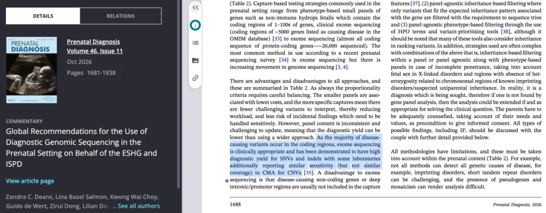
Figure. The 2026 joint recommendations of ESHG and ISPD state that whole-exome sequencing is suitable for prenatal clinical diagnosis, with a high diagnostic detection rate for SNV and InDel.
Why Do Ultrasound Anomalies Remain Unexplained by Current Testing?
The etiology of fetal structural anomalies is highly complex and may involve chromosomal abnormalities, single-gene variants, environmental factors, and more. Different testing technologies target different genetic levels:
Chromosomal karyotype analysis primarily detects chromosome number abnormalities and large structural rearrangements;
CMA primarily detects chromosomal copy number variations (CNVs);
WES focuses on single-gene-level abnormalities, including single nucleotide variants (SNVs), small insertions/deletions (Indels), and small copy number variations (CNVs).
Therefore, normal karyotype and CMA results do not completely rule out a genetic etiology. For fetuses with significant structural anomalies, particularly those suspected of having monogenic disorders, WES may help further identify the causative variant.
How Much Additional Diagnostic Yield Can WES Provide?
The PAGE study, published in The Lancet in 2019, enrolled 610 fetal cases with structural anomalies detected by ultrasound, in which aneuploidy and large CNVs had already been excluded, and used WES to further investigate the underlying genetic etiology[2]. The results showed:
An overall diagnostic yield of 8.5% (52/610);
A diagnostic yield of 15.4% in fetuses with multi-system structural anomalies;
A diagnostic yield of 15.4% in fetuses with skeletal abnormalities;
A diagnostic yield of 11.1% in fetuses with cardiac abnormalities.
The study demonstrated that WES can identify pathogenic variants that conventional chromosomal testing fails to detect, with particularly high diagnostic value in cases of multi-system anomalies and skeletal dysplasia. For prenatal diagnosis, establishing a genetic etiology not only explains why the fetus presents with an anomaly, but may also inform subsequent clinical decisions such as prognosis assessment, genetic counseling, and perinatal management.
AIExome® Human Exome Panel V5-Inherit: Focus on Genetic Disease Detection, Empowering Precision Diagnosis through Technological Innovation
Given the complex and diverse demands of genetic disease testing, comprehensive and accurate genomic information is a critical foundation for improving diagnostic capability. Building on more than a decade of expertise in gene capture technology, iGeneTech has launched AIExome® Human Exome Panel V5-Inherit, which, through optimized target region design and an enhanced capture system, provides more comprehensive technical support for genetic disease screening, prenatal genetic counseling, and research.
1. A Three-Tier Defense Network for CNV Detection
To address the limitations of conventional whole-exome sequencing in copy number variation (CNVs) detection, AIExome® Human Exome Panel V5-Inherit innovatively establishes a three-tier CNV detection network that accommodates both fine-scale intragenic variants and larger whole-genome CNVs.

Figure. The multi-layer probe design strategy of AIExome V5-Inherit for CNV detection: combining a whole-genome backbone, denser coverage of frequently affected regions, and single-exon-level design to balance CNV detection breadth with resolution in key regions.
For intragenic CNV detection, AIExome® Human Exome Panel V5-Inherit applies gene-specific probe design to key genetic disease-related genes, optimizing coverage of exons and adjacent regions and effectively enhancing the detection of single-exon and multi-exon deletions, duplications, and other CNVs. Validation data of AIExome® Human Exome Panel V5-Inherit demonstrate effective identification of representative variants such as a single-exon deletion of the DMD gene (Exon 74), a multi-exon deletion (Exons 48–50), and the −α⁴.² deletion in α-thalassemia, providing technical support for the detection of CNVs associated with genetic disorders such as Duchenne muscular dystrophy and thalassemia.
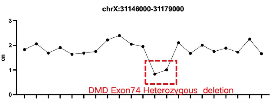
Figure. Example of heterozygous deletion of DMD Exon 74.

Figure. Example of heterozygous deletion of DMD Exons 48–50.

Figure. Example of −α⁴.² α-thalassemia deletion detection.
For regional and whole-genome CNV detection, AIExome® Human Exome Panel V5-Inherit provides high-density probe coverage of clinically relevant microdeletion regions such as 22q11.2, 1p36, and 17q12, introduces a whole-genome CNV backbone at 200 kb intervals, and offers optional 50/100 kb schemes, forming a multi-layered coverage system spanning from fine intragenic detection to the analysis of larger-scale CNVs.

Figure. Distribution of whole-genome SNP backbone loci: the number of SNPs and coverage across chromosomes is shown in 200 kb windows.
Through this three-tier CNV detection strategy, AIExome® Human Exome Panel V5-Inherit extends CNV detection capability beyond routine SNV and Indel detection, providing richer variant analysis information for genetic disease research and prenatal genetic diagnosis.
2. Accurate and Reliable Detection of SNV and InDel Variants
In terms of disease-related variant coverage, AIExome® Human Exome Panel V5-Inherit comprehensively covers clinically significant variant sites in the ClinVar database, supporting the identification and interpretation of genetic variants. Meanwhile, AIExome® Human Exome Panel V5-Inherit delivers robust SNV and Indel detection performance. Based on validation with the NA12878 reference sample, SNP detection achieves a maximum Precision of 99.71%, Recall of 99.74%, and F1-score of 99.72%.

Figure. Concordance analysis of variant calls using the NA12878 reference standard.
3. High-Depth Mitochondrial Sequencing Captures Heteroplasmic Variants
In addition, AIExome® Human Exome Panel V5-Inherit adopts full-length mitochondrial probe design, achieving high-depth coverage of mitochondrial DNA while maintaining autosomal sequencing performance, opening up more possibilities for variant analysis of mitochondria-related genetic disorders.
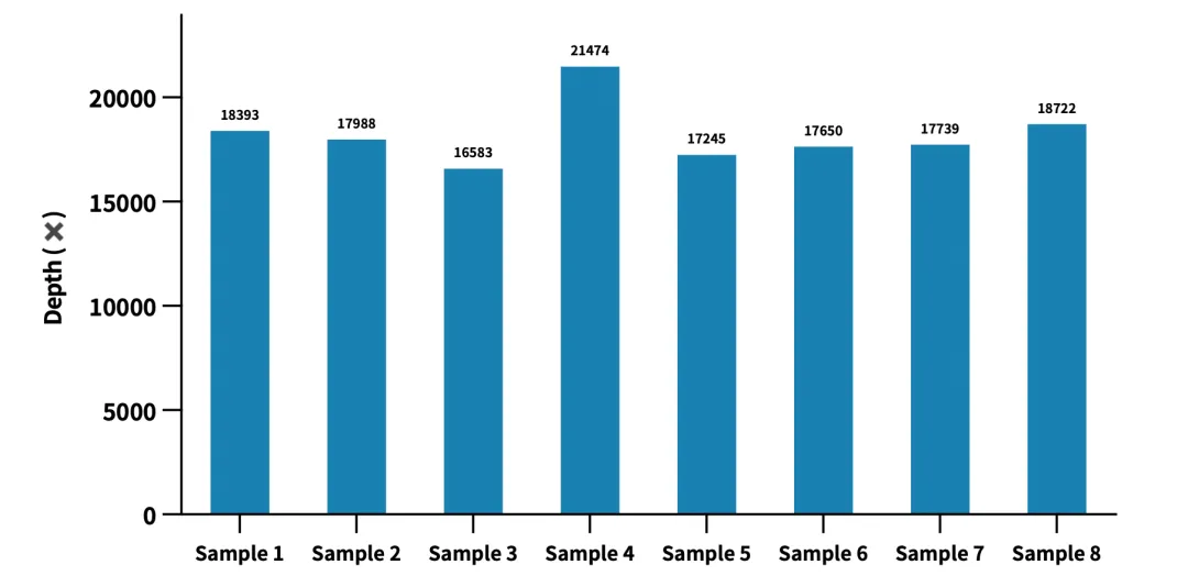
Figure. Assessment of mitochondrial sequencing depth based on real-world data of AIExome® Human Exome Panel V5-Inherit. Using a cell-line reference standard with enzymatic fragmentation library construction, 750 ng of input library, and overnight hybridization, the average mitochondrial depth consistently exceeds 10,000× at an average depth of 100× over the AIExome® V5-Inherit target regions.
From coding regions to key CNV regions and on to the mitochondrial genome, iGeneTech continues to refine the detection scope and technical performance of whole-exome capture, striving to minimize potential detection blind spots and provide a high-quality data foundation for prenatal genetic diagnosis and genetic disease research.
Exome capture is an important tool for prenatal genetic diagnosis, but different WES products vary considerably in gene coverage, capture efficiency, and coverage uniformity. Insufficient coverage of key genes may lead to missed pathogenic variants. In our next article, we will explore further: “Whole-Exome Sequencing ≠ Complete Coverage: Detection Blind Spots That Cannot Be Ignored in Prenatal Genetic Diagnosis,” analyzing the causes of coverage differences across WES assays and the key metrics for high-quality exome capture technology.
References
[1] Deans ZC, Basel Salmon L, Choy KW, et al. Global recommendations for the use of diagnostic genomic sequencing in the prenatal setting on behalf of the ESHG and ISPD. European Journal of Human Genetics. Published September 16, 2026. DOI: 10.1038/s41431-026-02217-4.
[2] Lord J, McMullan DJ, Eberhardt RY, et al. Prenatal exome sequencing analysis in fetal structural anomalies detected by ultrasonography (PAGE): a cohort study. The Lancet. 2019;393(10173):747–757. DOI: 10.1016/S0140-6736(18)31940-8.
 CN